See Inside Cells Like Never Before in These Vivid Microscopic Images
See Inside Cells Like Never Before in These Vivid Microscopic Images
以下正文同步自 Hacker News 最新,版权归原站所有,已转换为易读排版。
Home
See Inside Cells Like Never Before in These Vivid Microscopic Images
Comment
Save Article
Read Later
Share
Copied!
Copy link
Ycombinator
Comment
Comments
Save Article Read Later
Read Later
image gallery
See Inside Cells Like Never Before in These Vivid Microscopic Images
By
Molly Herring
October 21, 2026
Save Article
Read Later
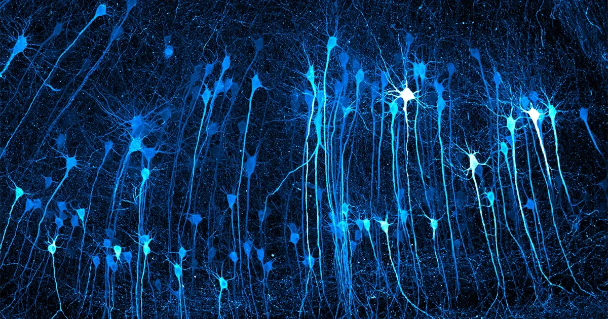
For this image of neurons in mouse brain tissue, Kaelin Wulf and Rachel Flannery of the Allen Institute used a home-built light-sheet microscope. In light-sheet fluorescence microscopy, a thin sheet of light illuminates a sample, which can then be scanned plane by plane. “I hope people can take away a sense of awe at how intricate, beautiful, and unimaginably vast the brain is,” Wulf said.
Kaelin Wulf, Rachel Flannery/Allen Institute; all photos courtesy of Nikon Small World
biology
cell biology
image gallery
All topics
On a frigid February morning in Edinburgh, Scotland, Patrick Hickey had run out of living things to put under his microscope. The plants and fungi in his garden had dried up, and summer puddles, with their flamboyant biological complexity, had long frozen over.
Hickey, a mycologist at Hypha Research Ltd., was using plant leaves to study mitochondria, cellular organelles that generate energy. He was planning to sprout mustard seeds for fresh samples, but he was impatient. While making coffee in his kitchen, he noticed a cold-hardy turtle vine hanging from the windowsill. “I snipped off an inch or so, put it in my pocket, took it into the lab, and was just amazed at what I saw,” Hickey said.
Under the microscope, mitochondria darted around the cells. They nimbly maneuvered around slower chloroplasts, like motorcyclists dodging traffic. “It was absolutely alive with activity,” Hickey said.
His time-lapse video of this microscopic dance, which leads our gallery below and was recently honored in Nikon’s 2026 Small World in Motion Competition, reveals the close partnership between the organelles: chloroplasts (red) spin sunlight into sugar, and mitochondria (green) convert sugar into energy.
The Nikon Small World competition, established in 1974, honors excellence in microscopy. The annual contests have charted not only the evolution of creative photomicrography but also the progress of imaging technology.
Hickey — and many of the other honorees featured here — relied on a technique called confocal laser scanning microscopy.
Unlike a standard microscope, which illuminates an entire sample at once and can produce blurry images, a confocal microscope focuses on one depth level at a time. Researchers first “label” a sample with fluorescent dyes that attach to specific structures within the cell. A laser then illuminates a thin layer of the sample, and the microscope detects the resulting fluorescence. Scanning line by line, the system builds a detailed image.
As imaging technology advances, so too does our understanding of cellular processes. Hickey’s video shows that mitochondria are more dynamic than previously thought, which suggests that they are “more than just a ‘powerhouse of the cell,’” he said. “They are likely involved in cytoplasmic streaming, signaling, and interactions with other organelles.”
The full list of awardees can be found here for the photo contest and here for the video contest. We’ve chosen to feature a several winners and honorable mentions that reveal the intricacies of the cellular world.
Mitochondria (green) navigate around chloroplasts (red) in the leaf cells of a turtle vine. “Their movement looked like rivers of electricity flowing within the cells,” Hickey said.
To study diseases, Gauri Kulkarni of Wake Forest University School of Medicine grows miniature organs in her lab, including the 1.3-millimeter human gut tissue shown in this image. The glowing network reveals the connections between epithelial cells that make up a healthy gut barrier. This barrier acts like a microscopic security guard, keeping harmful microbes and food toxins from leaking into the bloodstream. “In science, we often rely on cold data and numbers to understand disease, but photomicrography bridges the gap between logic and emotion,” Kulkarni said.
Gauri Kulkarni/Wake Forest University School of Medicine
This video shows how heart muscle cells contract and how calcium regulates their beating. The green chains are sarcomeres, the smallest contractile units of heart muscle. The red flashes show calcium inside the cell. At the start of a beat, an electrical impulse sends calcium into the cell, which triggers the release of more calcium. All this calcium tells the sarcomeres to contract. The calcium then leaves the cytoplasm, which allows the cell to relax in preparation for the next beat. “The same video can reveal the beauty of living heart cells while providing quantitative information about their structure and function,” said Yasaman Maaref of Simon Fraser University, who created the video.
This video of a mouse’s brain cells shows actin (blue), a protein that helps the cell change shape, and lysosomes (orange), organelles that break down cellular waste. While both structures move, the lysosomes are much faster, making the actin network appear almost stationary in comparison.
Bruno Cisterna, Halli Lindamood, Eric Vitriol/Medical College of Georgia at Augusta University
The top layer of this image of a mouse trachea shows the epithelium, the outer layer of skin cells. Each green dot is the nucleus of a single cell. Beneath, a layer of black basement membrane, mostly made of collagen, provides structure and acts as a filter. Next, a thick layer of yellow reveals the actin packed inside smooth muscle cells that control the movement of air through the trachea. And that faint layer of dark blue at upper right? That’s “the kind of mucus you cough and spit out,” said Julien Resseguier, who created the image to study the interplay between neural and immune systems.
This image of a rat liver endothelial cell shows actin filaments (brownish orange) and microtubules (white). Both of these protein structures work together to give the cell its shape. Intracellular gaps are visible in the image; these holes can disrupt the liver’s protective blood barrier and might indicate injury, inflammation, or cancer.
Francisco Lázaro-Diéguez/Albert Einstein College of Medicine
Embryonic cells from the skin of Xenopus, a clawed frog, have cilia, seen here in green, that beat in unison to generate a wave of fluid movement past the cell surface. Confocal laser scanning microscopy allowed the researchers to distinguish individual cilia, the centrioles that anchor them (red), and cell boundaries (white) that help the cells stick together.
Pierre Nouaux, Laurent Kodjabachianan, Camille Boutin/Center for Integrative Biology in Toulouse
Trypanosoma cruzi, the parasite that causes Chagas disease, is an intracellular pathogen that can infect almost all mammal cell types. Richard Atherton of the London School of Hygiene and Tropical Medicine captured this image of a T. cruzi nest inside a host cell. “If my image can raise even a little more awareness about Chagas disease, then I will be happy,” Atherton said. “I also hope my image can get people excited about science, nature, and the infinite microscopic worlds hidden all around us.”
Richard Atherton/London School of Hygiene and Tropical Medicine
This video, by Andrew Moore of the Howard Hughes Medical Institute, illuminates how actin (white) and mitochondria (blue) interact during cell division. Moore found that the actin seemed to be involved in positioning and organizing the mitochondria; the actin cycles through the mitochondrial network in a wave and forms “comet tails” that drive bursts of motion. Over the course of mitosis, these comet tails “mix up the mitochondrial network, shuffling their spatial position, almost like a card dealer shuffling a deck of cards,” Moore said.
Andrew Moore/Howard Hughes Medical Institute
Get highlights of the most important news delivered to your email inbox
Save Article
Read Later
How Does Life Unfold? A Landscape Metaphor Comes Into Its Own.
development
How Does Life Unfold? A Landscape Metaphor Comes Into Its Own.
By
Philip Ball
October 9, 2026
Comment
Save Article
Read Later
Surprisingly Complex Waves Reveal the Brain’s Inner Workings
neuroscience
Surprisingly Complex Waves Reveal the Brain’s Inner Workings
By
Conor Feehly
September 30, 2026
Comment
Save Article
Read Later
Comment on this article
Quanta Magazine moderates comments to facilitate an informed, substantive, civil conversation. Abusive, profane, self-promotional, misleading, incoherent or off-topic comments will be rejected. Moderators are staffed during regular business hours (New York time) and can only accept comments written in English.
正文由 探How 从来源站点 RSS 同步,内容未经改写;遇到排版缺失或需要图片、视频时请以原文为准。