Action Potentials for October
1: Blood flow in the brain is incredibly important for basically everything. Beyond dementia, wherein impairments in blood flow are a major risk factor, regional impairments in blood circulation have also been associate…
以下正文同步自 Hacker News 最新,版权归原站所有,已转换为易读排版。
Andy McKenzie
Oct 31, 2026
5
1
Share
1: Blood flow in the brain is incredibly important for basically everything. Beyond dementia, wherein impairments in blood flow are a major risk factor, regional impairments in blood circulation have also been associated with plenty of psychiatric disorders over the years, such as some subtypes of depression.
Anatomically, the major arteries and veins travel across the pial surface of the brain and then penetrate into the parenchyma at approximately perpendicular angles:
visualizing non-capillary vessels only
A new study re-analyzes the blood vessels from a previously published connectomics data set of one cubic millimeter of mouse visual cortex, which was preserved by aldehyde fixation and imaged via electron microscopy.
Here is the capillary network, which they defined based on the topology of the vascular network. Most of these capillary vessels are 4-5 μm in diameter:
n = 7757 of the 9292 total capillaries, colored based on their branch order from the blood source (J) or blood drain (K)
Other than these vascular lumen, they classified the other structures in the volume into six categories: (a) thin axons, (b) “perivascular” structures — i.e. endothelial cells and mural cells, (c) dendrites, (d) glia, (e) neuronal cell bodies (somata), and (f) thick or myelinated axons. Separately, they used synapse annotations, which were considered sub-parts of the dendrites and axons. Here’s an example of these structures around one blood vessel:
https://www.biorxiv.org/content/10.64898/2026.09.26.754479v1
Once they did all these classifications, they got to the surprising part of the paper, which is how these structures associated with one another.
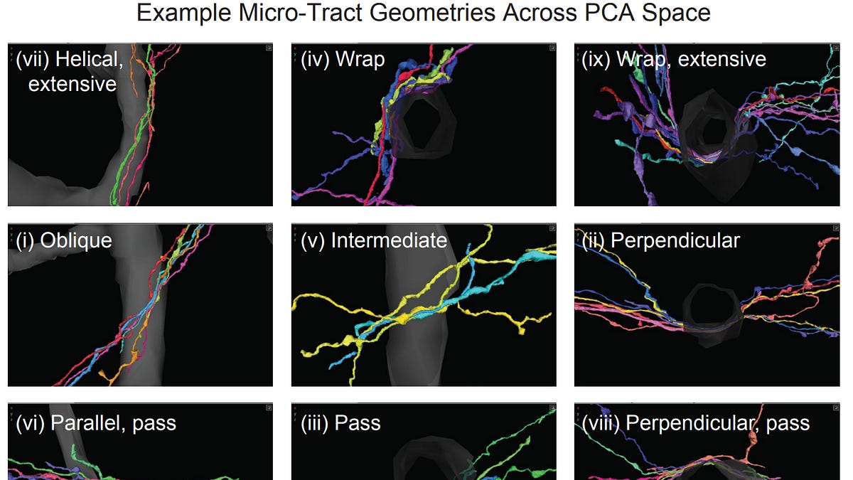
Specifically, they found that axons (but not dendrites) tend to form micro-tracts running alongside blood vessels:
example axon microtracts; axons = colored, blood vessel = grey
These micro-tracts can form at different geometric angles relative to the blood vessels:
https://www.biorxiv.org/content/10.64898/2026.09.26.754479v1.full.pdf
Why does this matter? The authors speculate that it might be a mechanism for neurovascular coupling, whereby when there is more action potential firing, the local concentration of potassium outside of the axons might increase, and this could be picked up by cells in the blood vessel and lead to changes in blood vessel diameter. But mostly, it’s just an interesting anatomical finding that awaits future understanding.
2: 2D images, such as those produced by Golgi-Cox impregnation or confocal microscopy of fluorescent neurons, are what have been used to classify the anatomy of dendritic spines for more than 100 years.
But a new study uses 3D connectomics data to show how such 2D images can be misleading about what type of structure is present. For example, when they rotate one dendritic shaft, spines that would have been classified as long or mushroom based on Angle 1 instead look stubby on Angle 2:
https://journals.physiology.org/doi/full/10.1152/jn.00280.2026
3: Synaptopodin is a protein that binds actin and affects the organization of the endoplasmic reticulum and thus calcium signaling. Mice that lack synaptopodin have deficits in long term potentiation (LTP) and learning.
A new study creates a genetically modified version of rats that lack synaptopodin. Structurally, they find that such rats lack a spine apparatus in dendrites and lack a cisternal organelle in axon initial segments, which are both forms of specialized endoplasmic reticulum.
Notably, these rats had normal numbers of excitatory synapses and a normal thickness of their hippocampal CA1 layers. However, they did have deficits in LTP. This may be an example of how alterations in intracellular organization can affect neural signaling.
red = postsynaptic density, green = spine apparatus, which is absent in rats lacking synaptopodin (KO)
green = smooth endoplasmic reticulum, magenta = cisternal organelles, blue = symmetric synapses
4: Although we don’t always think about it in this way, your brain is a physical object sitting inside of your skull, protected by dura mater (unless, of course, you are an AI).
The tentorium is the dura that separates the cerebrum from the cerebellum. And where the tentorium abuts into the medial temporal lobe, it often produces an indentation, which is called the tentorial notch (also known as the uncal notch):
https://alz-journals.onlinelibrary.wiley.com/doi/10.1002/alz.71827
A new study finds that although the tentorial notch is very common, it varies substantially in severity between people:
https://link.springer.com/article/10.1186/s40478-026-02414-8
Interestingly they also report that a more severe tentorial notch is associated with cell damage. Here I am a little skeptical. Their definition of “cell damage” includes when neurons look stretched or the cortical layers are displaced, but to me those don’t necessarily indicate that there would be significant problems in how the neuronal network would function.
https://link.springer.com/article/10.1186/s40478-026-02414-8
5: Speaking of skull shape, this past month the NYT profiled my research residency mentor (and Apex Neuroscience advisor) John Crary for his research relating head shape to risk of brain pathology (such as tau accumulation).
Somewhat oddly to me the hook was “it’s not phrenology,” but I don’t think anyone was thinking that? But hey, I understand that finding everyday hooks for neuroscience content is tricky (which is why I myself don’t even try, I just write only for my fellow brain nerds 🤓).
Although the particular data that they discussed in the NYT article is not necessarily the strongest available in the whole field, I personally find John’s overall hypothesis that a history of subtle mechanical stresses which accumulate over an ordinary lifetime could explain why either (a) certain regions (e.g. in the entorhinal cortex, because it is exposed to the skull base) or (b) certain people develop tau pathology sooner than others to be extremely plausible.
6: A new autopsy study from the University of Sao Paulo presents a large (n = 2711) data set correlating cognitive impairment with neuropathologic and demographic variables. The average age was 73.4 with a standard deviation of 14.4 years and a range of 23 to 111.
They used a statistical method (LMG) to find out how much each variable contributes to the variance explained by a regression model.
They found that Braak stage (i.e. the distribution of tau pathology) was the strongest predictor of cognitive impairment, followed by “contributing coexisting pathology” (cerebrovascular disease, Lewy body pathology, and TDP-43 pathology — unfortunately they didn’t distinguish these), then Thal phase and CERAD score (measures of amyloid pathology), then age, and then sex.
data from https://www.medrxiv.org/content/10.64898/2026.10.20.26365945v1.full.pdf
One way to interpret this is that we have gotten pretty good at using neuropathology to predict whether elderly people will have cognitive impairment, so much so that age itself is not as strong a predictor as those neuropathologic measures. But unfortunately, while we have good associations, it’s not clear the extent to which these are causal or they are marking for something elsewhere. I think this is the main reason that our treatments or preventative measures for dementia are not very good yet.
7: I remember a few years ago I was reading about the circulating DNA blood test for early cancer detection. At the time I wondered whether I should try to sign my parents up for it. It was expensive, and I wasn’t sure they’d qualify, so I didn’t look into it very much, but I did some basic research on it.
In February, the Galleri blood test reported non-significant results from its trial, and last month a paper was published on it. It was a huge study of 142,250 people, with half randomized to get the blood test and half not. Unfortunately, it didn’t meet the endpoint — there was no significant reduction in combined stage III and IV cancers in 12 specified cancer types.
Much is being made of the pre-specified secondary endpoint of a lower incidence of stage IV cancers in the intervention group, but it’s barely nominally significant (95% CI: 0.744–0.998), and I’m generally skeptical of most secondary endpoints in trials like this.
One of the key problems seems to be that it has poor sensitivity. There are still a ton of open questions in the field and I still expect this to improve and be useful at some point. However, I also interpret this result as weak Bayesian evidence for the idea that early cancer detection is hard/doesn’t help that much for some of the deadliest cancers, because the really deadly cancers grow so aggressively that it’s really hard to intervene on them anyway.
8: Charles River stock has been up a lot this past month, potentially as a “picks and shovels” idea on the newfound interest in AI x Bio investment. Their business model seems to be primarily contract research, much of which involves experiments on rodents and nonhuman primates.
However, it’s still down from its highs in 2021, when biotech was really ripping:
9: A new connectomics data set of the central nervous system of a first instar (i.e. the earliest larval developmental stage) female fruit fly.
One thing I find interesting about this is that — to the best of my understanding — the data was imaged in 2018, but these automated segmentations are being released only now, 8 years later. Although people expect computational advances to dramatically accelerate connectomics in the future, at least for now, it seems like computational bottlenecks in reconstructing neural circuits are still very much a thing. The authors estimate that fully proofreading the reconstructions with current methods would require “either tens of man-years of effort or significantly improved superpixel agglomeration methods, yet to be developed by us or others.”
https://www.biorxiv.org/content/10.64898/2026.09.28.754924v1.full.pdf
10: A new study performs spatial transcriptomics on human brain tissue that had been fixed for between 1 month to 2.2 years. They found that with more aggressive target retrieval (longer heat treatment, to reverse the crosslinks), they could still obtain useful transcriptomic data from the long-term fixed tissue, although longer fixation was still associated with less RNA detection. In my opinion this is most likely due to worse RNA accessibility to the probes, due to prolonged formaldehyde crosslinking.
https://pmc.ncbi.nlm.nih.gov/articles/PMC13372838/
11: Preclinical paper on a new preventative treatment for prion disease finds that it is effective at improving survival in a mouse model. I remember reading about Sonia Vallabh and Eric Minikel’s story back in the mid-2010s, and their efforts since then have definitely lived up to their lofty goals (e.g., they are the senior authors on this paper). Most impressive.
https://www.biorxiv.org/content/10.64898/2026.10.22.761034v1
12: Kevin Kennedy argues that the efficacy in large trials of TMS for depression is much weaker than commonly recognized, especially when you look at the larger clinical trials (which, of course, are the more reliable ones).
13: Expanse Bio LLC received a new grant from ARPA-H with up to $14.2 million in funding. Dr. Sebastian Giwa is the Principal Investigator. Their goal is to develop vitrification techniques to bank “viable” brain tissue.
As always, viability is a confusing word in cryobiology, having long meant different things in different contexts. It can refer to cell membrane integrity, various types of metabolic activity, (some degree of) cell survival or ability to proliferate, maintenance of tissue-specific function such as electrophysiology, or organ functioning after transplantation (this last meaning is, naturally, how it is typically used in the transplant world). I’d be very curious to hear what viability e
正文由 探How 从来源站点 RSS 同步,内容未经改写;遇到排版缺失或需要图片、视频时请以原文为准。